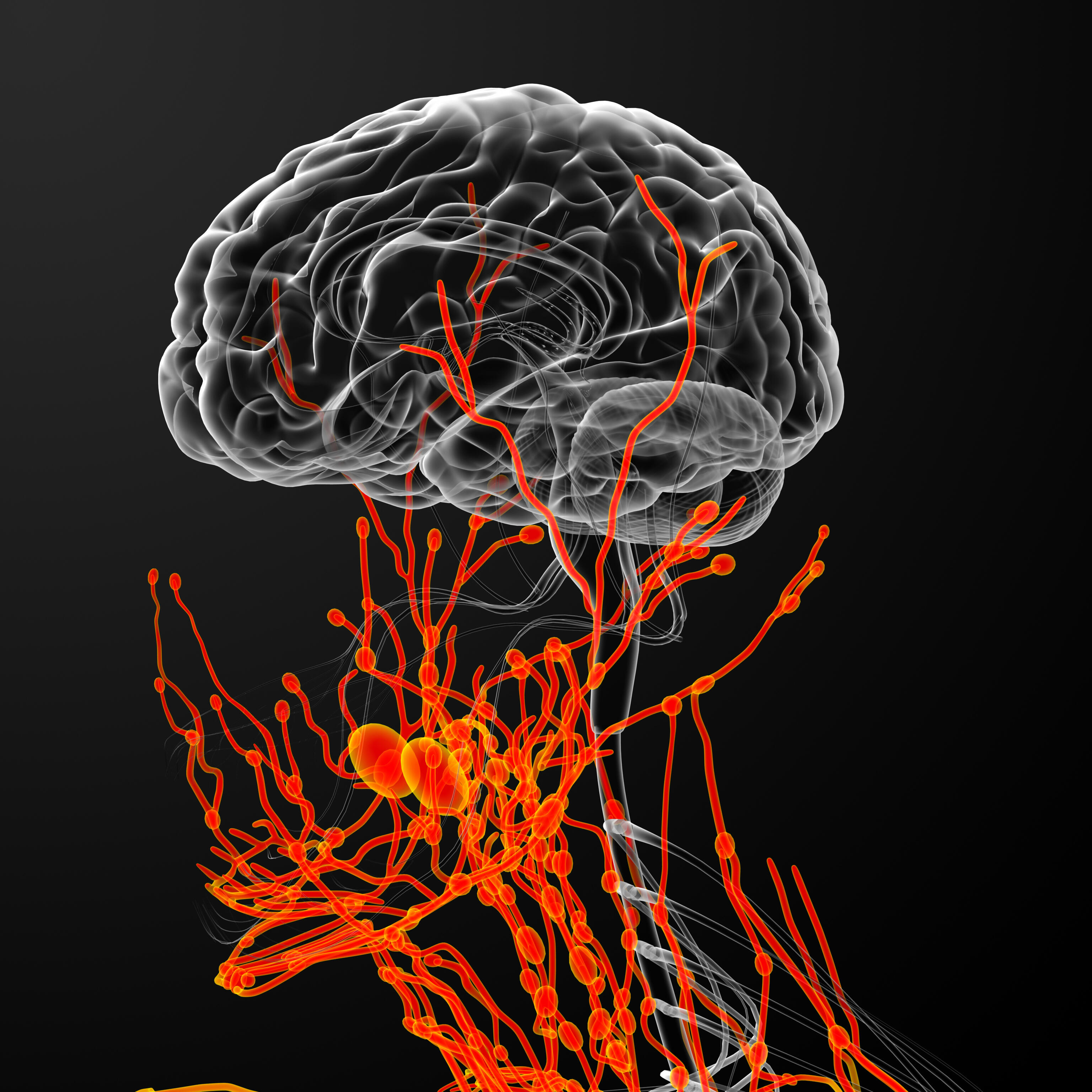
Amyloid-beta (Aβ) accumulation in Alzheimer’s Disease (AD) may, in part, be due to an impairment in the brain’s clearance mechanisms, which would normally remove waste proteins from the interstitial fluid surrounding neurons or from the cerebrospinal fluid. Strategies promoting clearance and preventing the build-up of Aβ may be useful therapeutically – but first we need to understand what to target.
The accumulation of amyloid-beta (Aβ) into parenchymal plaques or in the walls of cerebral capillaries and arteries (cerebral amyloid angiopathy) are characteristic pathologic features of Alzheimer’s Disease (AD). This build-up may result from an imbalance between the production and clearance of Aβ,.1
In the brain, soluble proteins such as Aβ can be removed or ‘cleared’ from the interstitial fluid (ISF) around neurons or from the cerebrospinal fluid (CSF) via a number of hypothesized pathways.1 These pathways may offer potential targets for the treatment of AD, by promoting clearance and preventing the build-up of Aβ or other neurotoxic proteins. In this symposium, four speakers explored the mechanisms of interstitial fluid clearance.
IPAD may hold clues to cerebral amyloid angiopathy
Roxana Carare MD, PhD (University of Southampton, United Kingdom) considered the role of the glymphatic system in the accumulation of Aβ in cerebral arteries. Imaging studies in mice suggest that ISF is cleared from the brain tissue by passing into the basement membranes of capillaries, flowing through cerebral arteries towards lymph nodes in the neck, before exiting through the basement membranes of the arteries – a process called the intramural peri-arterial drainage (IPAD) pathway.2
Intramural peri-arterial drainage may offer a potential therapeutic target
A recent study in mice showed that Aβ from the CSF can enter the brain through the basement membranes of cortical arteries and leave via the IPAD pathway.2 In older mice however, Aβ can be seen in astrocytes and macrophages in the parenchyma as well as the capillary basement membrane – indicating a possible defect in the IPAD pathway. The IPAD pathway may therefore offer a potential therapeutic target for the future.
Characteristics of Enlarged Perivascular Spaces in Alzheimer’s Disease
Joel Ramirez, PhD (Sunnybrook Research Institute, University of Toronto, Canada) reviewed the use of neuroimaging to study enlarged perivascular spaces (ePVS) in the brains of people with AD. Waste products, including Aβ can accumulate around blood vessels in the brain and may cause blockages in the perivascular space and prevent ISF drainage. As a result the
perivascular space may become enlarged. ePVS, some >3 mm, can be seen using a number of MRI imaging techniques, and are markers of small vessel disease in the brain.3, 4 In addition, the location of the ePVS seems to reflect the type of small vessel disease.5
The perivascular space may become enlarged
Until recently, quantitative estimates of ePVS relied on visual counts from MRI, however new automated methods using segmentation of images have been developed that could prove valuable tools in future clinical trials.6, 7 Using such a technique, the Sunnybrook Dementia Study showed that men have more ePVS in their white matter compared with women, and individuals with AD had more ePVS than unaffected normal elderly controls.5
Mouse studies suggest that ISF clearance occurs during sleep
Mouse studies suggest that ISF clearance occurs during sleep. In a study of 26 patients with suspected cerebrovascular disease, decreased slow-wave sleep and increased nocturnal arousals correlated with increased ePVS, suggesting that sleep may play a role in ISF drainage in humans too.8
Meningeal lymphatics in Aβ clearance
Natalia Acosta, MSc (University of Eastern Finland, Finland) presented recent data on the role of meningeal lymphatics in Aβ clearance. CSF has been hypothesized to drain through the meningeal lymphatic system. In transgenic mice that lack dural lymphatic vessels, CSF was not drained from the subarachnoid space and the transport of macromolecules was significantly reduced.9
Recent mouse studies have shown that a lack of meningeal lymphatics induces slight decreases in soluble cortical Aβ levels and hippocampal amyloid plaque load. Although mechanical clearance rates do not appear to increase, at the hippocampus the blood–brain barrier (BBB) appears to be ‘leaky’ and there is an increase in the number of T-cells. These findings suggest that the hippocampal Aβ reduction may not be the result of bulk fluid clearance.
Hippocampal Aβ reduction may not be the result of bulk fluid clearance
Link between blood–brain barrier dysfunction and Alzheimer’s Disease
Berislav Zlokovic, Ph.D. (Zilkha Neurogenetic Institute, University of Southern California, USA) examined the link between the breakdown of the BBB and the development of AD.
Not only do genetic defects in a number of BBB transporters and cellular junctions lead to neurologic disorders, but many genes underlying neurodegenerative diseases are associated with BBB breakdown and cerebrovascular dysfunction.10 One example is the PICALM gene, which plays a role in Aβ clearance across the BBB; endothelial cells carrying the protective allele showed greater expression of PICALM and improved Aβ clearance, compared with cells carrying the non-protective allele.11 A new study is currently modelling eight new PICALM AD mutations.
In sporadic AD, BBB breakdown occurs as a result of pericyte degeneration allowing an influx of toxic proteins from the blood into the brain; interestingly, this process is accelerated by apolipoprotein E4 but may be independent of Aβ.12 New data in mouse models show that although the presence of Aβ42 or tau does not alter BBB permeability, permeability does increase with cognitive impairment. In addition, APOE4 carriers appear to have greater BBB permeability in the hippocampus compared with APOE3 carriers, which is further increased in those with mild cognitive impairment.
Ongoing clinical trials are now targeting the BBB and pericytes
A number of ongoing clinical trials are now targeting the BBB and pericytes in a number of neurodegenerative diseases. Strategies include blocking RAGE (receptor for advanced glycation end products) to seal the BBB, preventing Aβ accumulation, and protecting against vascular damage and inflammation.